Shoulder Dislocations: A Comprehensive Guide
What it is:
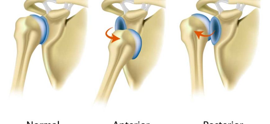
A shoulder dislocation occurs when the head of the upper arm bone (humerus) is forced completely out of its socket (glenoid), which is part of the shoulder blade (scapula). The shoulder is the body’s most mobile joint, but this great range of motion comes at the cost of stability, making it the most commonly dislocated major joint in the body.
Types:
Dislocations are classified by the direction in which the humeral head moves and by whether it is a first-time or recurring injury.
- Anterior Dislocation: The most common type (over 95% of cases). The arm bone is pushed forward, out of the socket. This often happens when the arm is forced into an extreme position, like during a throwing motion or a fall on an outstretched hand.
- Posterior Dislocation: Much rarer (2-4% of cases). The arm bone is pushed backward out of the socket. This can be caused by a direct blow to the front of the shoulder, a seizure, or an electric shock.
- Inferior Dislocation (Luxatio Erecta): Very rare. The arm bone is displaced downward, leaving the arm fixed in an overhead position. This requires significant force.
- Recurrent Dislocation: After an initial dislocation, the supporting structures often stretch or tear, making the shoulder prone to dislocate again with minimal force. This is especially common in young, active patients.
Symptoms:
- Intense, severe pain in the shoulder and upper arm.
- A visible deformity or “bump” (the humeral head) in front of or behind the normal shoulder contour.
- Swelling and bruising.
- Inability to move the arm from its awkward position.
- Numbness, tingling, or weakness in the arm, neck, or hand (a sign of potential nerve or blood vessel impingement).
Diagnosis:
- Physical Examination: A doctor will assess the deformity, check for areas of numbness, and evaluate pulses to ensure no vascular injury.
- X-rays: Essential and required before and after treatment. They confirm the dislocation, show the direction, and check for associated fractures (which occur in a significant number of dislocations).
- MRI: Not typically used for the initial diagnosis but is extremely valuable afterward to assess damage to the soft tissues, especially the labrum (a ring of cartilage that deepens the socket) and rotator cuff tendons.
Prevention:
- Strength Training: Regularly strengthening the rotator cuff and scapular stabilizer muscles is the best prevention.
- Proper Technique: Using correct form in sports and weightlifting.
- Protective Gear: For athletes in contact sports with a history of dislocation, a brace may be used to prevent the arm from moving into vulnerable positions.
- Avoid Risky Positions: For those with previous dislocations, avoiding arm positions that have caused dislocation in the past (e.g., excessive overhead rotation).
Treatment:
- Immediate (Reduction): The dislocated bone must be put back into the socket, a procedure called closed reduction. This should be done by a trained medical professional with sedation or pain medication. Never try to pop it back in yourself, as this can cause further damage.
- Post-Reduction: After the joint is reduced, the arm is placed in a sling or shoulder immobilizer for comfort and to allow the soft tissues to begin healing. Ice and anti-inflammatory medication (e.g., ibuprofen) help manage pain and swelling.
- Physical Therapy: The cornerstone of non-surgical treatment. Therapy focuses on restoring range of motion and, most importantly, strengthening the muscles around the shoulder to improve dynamic stability and prevent recurrence.
Types of Surgeries:
Surgery is considered for first-time dislocations in young athletes or after recurrent dislocations have occurred.
- Arthroscopic Bankart Repair: The most common procedure for anterior dislocations. Using a small camera and instruments, the surgeon reattaches the torn labrum and ligaments to the socket rim. This is a minimally invasive procedure.
- Latarjet Procedure: A more robust bone-block procedure used for significant bone loss on either the socket (glenoid) or the ball (humeral head). A piece of bone with an attached tendon is moved to the front of the glenoid to prevent the humerus from slipping forward.
- Open Stabilization: Less common now with advanced arthroscopy, but may be used for complex revisions.
Prognosis:
- Non-Surgical: The prognosis varies greatly by age. For older patients (over 40), the risk of recurrence is lower, but the risk of a concomitant rotator cuff tear is higher. For young athletes (under 25), the risk of recurrence without surgery can be over 80%.
- Surgical: Surgical stabilization has a very high success rate (90-95%+) in preventing future dislocations and allowing a return to sports. Recovery involves a period of strict immobilization followed by a lengthy and structured physical therapy protocol lasting several months.
Warning Signs & When to See a Doctor:
A shoulder dislocation is a medical emergency. You must go to the emergency room or an urgent care center immediately.
Go to the Emergency Room immediately. Additionally, be aware of these red flags after a reduction:
- Increased numbness or tingling in the arm or hand.
- The arm becomes increasingly pale, blue, or cold.
- Severe, unrelenting pain.
These are signs of potential nerve or vascular injury that require urgent attention.